|
Addgene inc
cj pet24a kan  Cj Pet24a Kan, supplied by Addgene inc, used in various techniques. Bioz Stars score: 90/100, based on 1 PubMed citations. ZERO BIAS - scores, article reviews, protocol conditions and more https://www.bioz.com/product/t7+expression+vector+pet-24a/T7-PglC-His6+(Plasmid+%2389709)/pmc06710627-467-72-38 Average 90 stars, based on 1 article reviews
cj pet24a kan - by Bioz Stars,
2026-09
90/100 stars
|
Buy from Supplier |
|
Addgene inc
cj pet 24a kan  Cj Pet 24a Kan, supplied by Addgene inc, used in various techniques. Bioz Stars score: 90/100, based on 1 PubMed citations. ZERO BIAS - scores, article reviews, protocol conditions and more https://www.bioz.com/product/t7+expression+vector+pet-24a/T7-PglD-His6+(Plasmid+%2389710)/pmc06710627-467-86-38 Average 90 stars, based on 1 article reviews
cj pet 24a kan - by Bioz Stars,
2026-09
90/100 stars
|
Buy from Supplier |
|
Addgene inc
pet30b kan  Pet30b Kan, supplied by Addgene inc, used in various techniques. Bioz Stars score: 91/100, based on 1 PubMed citations. ZERO BIAS - scores, article reviews, protocol conditions and more https://www.bioz.com/product/t7+expression+vector+pet-24a/pET30b(%2B)+mKO2-HuR%2Ewt+(Plasmid+%23110400)/pmc06710627-467-60-38 Average 91 stars, based on 1 article reviews
pet30b kan - by Bioz Stars,
2026-09
91/100 stars
|
Buy from Supplier |
|
Addgene inc
report 89723 Report 89723, supplied by Addgene inc, used in various techniques. Bioz Stars score: 90/100, based on 1 PubMed citations. ZERO BIAS - scores, article reviews, protocol conditions and more https://www.bioz.com/product/t7+expression+vector+pet-24a/T7-PseB-His6+(Plasmid+%2389723)/pmc06710627-467-93-38 Average 90 stars, based on 1 article reviews
report 89723 - by Bioz Stars,
2026-09
90/100 stars
|
Buy from Supplier |
|
Addgene inc
caption a7 construct vector resistance reference addgene id gst pglfδ1  Caption A7 Construct Vector Resistance Reference Addgene Id Gst Pglfδ1, supplied by Addgene inc, used in various techniques. Bioz Stars score: 91/100, based on 1 PubMed citations. ZERO BIAS - scores, article reviews, protocol conditions and more https://www.bioz.com/product/t7+expression+vector+pet-24a/GST-PglF130+(Plasmid+%2389708)/pmc06710627-480-32-38 Average 91 stars, based on 1 article reviews
caption a7 construct vector resistance reference addgene id gst pglfδ1 - by Bioz Stars,
2026-09
91/100 stars
|
Buy from Supplier |
|
GenScript corporation
truncated mouse papillomavirus l1 dna sequence Truncated Mouse Papillomavirus L1 Dna Sequence, supplied by GenScript corporation, used in various techniques. Bioz Stars score: 90/100, based on 1 PubMed citations. ZERO BIAS - scores, article reviews, protocol conditions and more https://www.bioz.com/product/t7+expression+vector+pet-24a/truncated+mouse+papillomavirus+l1+dna+sequence/us11858964-741-10-37 Average 90 stars, based on 1 article reviews
truncated mouse papillomavirus l1 dna sequence - by Bioz Stars,
2026-09
90/100 stars
|
Buy from Supplier |
|
Cytiva Europe
histrap hp column Histrap Hp Column, supplied by Cytiva Europe, used in various techniques. Bioz Stars score: 97/100, based on 1 PubMed citations. ZERO BIAS - scores, article reviews, protocol conditions and more https://www.bioz.com/product/t7+expression+vector+pet-24a/HisTrap+HP/pmc05137464-137-93-96 Average 97 stars, based on 1 article reviews
histrap hp column - by Bioz Stars,
2026-09
97/100 stars
|
Buy from Supplier |
|
Cytiva Europe
äkta start system äkta Start System, supplied by Cytiva Europe, used in various techniques. Bioz Stars score: 96/100, based on 1 PubMed citations. ZERO BIAS - scores, article reviews, protocol conditions and more https://www.bioz.com/product/t7+expression+vector+pet-24a/%C3%84KTA+start/pmc05137464-137-79-82 Average 96 stars, based on 1 article reviews
äkta start system - by Bioz Stars,
2026-09
96/100 stars
|
Buy from Supplier |
|
Promega
a purified ecorv-restricted pet24a( ) vector A Purified Ecorv Restricted Pet24a( ) Vector, supplied by Promega, used in various techniques. Bioz Stars score: 90/100, based on 1 PubMed citations. ZERO BIAS - scores, article reviews, protocol conditions and more https://www.bioz.com/product/t7+expression+vector+pet-24a/pgem+t+easy+plasmid/pm17921261-51-8-11 Average 90 stars, based on 1 article reviews
a purified ecorv-restricted pet24a( ) vector - by Bioz Stars,
2026-09
90/100 stars
|
Buy from Supplier |
|
Addgene inc
pex3 amino acids 1 42 fkbp mcherry fk506 binding protein domain fkbp fragment Pex3 Amino Acids 1 42 Fkbp Mcherry Fk506 Binding Protein Domain Fkbp Fragment, supplied by Addgene inc, used in various techniques. Bioz Stars score: 92/100, based on 1 PubMed citations. ZERO BIAS - scores, article reviews, protocol conditions and more https://www.bioz.com/product/t7+expression+vector+pet-24a/pIRES-Ap2a2-FKBP+(Plasmid+%2346944)/bio_rxiv__2023__08__21__554162-211-24-44 Average 92 stars, based on 1 article reviews
pex3 amino acids 1 42 fkbp mcherry fk506 binding protein domain fkbp fragment - by Bioz Stars,
2026-09
92/100 stars
|
Buy from Supplier |
|
Addgene inc
vector pegfp c1 Vector Pegfp C1, supplied by Addgene inc, used in various techniques. Bioz Stars score: 93/100, based on 1 PubMed citations. ZERO BIAS - scores, article reviews, protocol conditions and more https://www.bioz.com/product/t7+expression+vector+pet-24a/pMito-mCherry-FRB+(Plasmid+%2359352)/pmc10830484-337-42-49 Average 93 stars, based on 1 article reviews
vector pegfp c1 - by Bioz Stars,
2026-09
93/100 stars
|
Buy from Supplier |
|
Addgene inc
mito mscarlet dennd6a  Mito Mscarlet Dennd6a, supplied by Addgene inc, used in various techniques. Bioz Stars score: 95/100, based on 1 PubMed citations. ZERO BIAS - scores, article reviews, protocol conditions and more https://www.bioz.com/product/t7+expression+vector+pet-24a/pmScarlet-i_C1+(Plasmid+%2385044)/bio_rxiv__2023__08__21__554162-211-9-14 Average 95 stars, based on 1 article reviews
mito mscarlet dennd6a - by Bioz Stars,
2026-09
95/100 stars
|
Buy from Supplier |
Image Search Results
Journal: Methods in enzymology
Article Title: Chemoenzymatic Synthesis and Applications of Prokaryote-Specific UDP-Sugars
doi: 10.1016/bs.mie.2017.06.003
Figure Lengend Snippet: Plasmids used in this manuscript.
Article Snippet: Transform plasmid into freshly-competent BL21 (DE3) RIL cells, growing transformants on LB-agar selection media containing corresponding vector selection antibiotic (see ) and 30 μg/mL CAM. table ft1 table-wrap mode="anchored" t5 Table 1. caption a7 Construct Vector Resistance Reference
Techniques: Plasmid Preparation, Modification
Journal: Methods in enzymology
Article Title: Chemoenzymatic Synthesis and Applications of Prokaryote-Specific UDP-Sugars
doi: 10.1016/bs.mie.2017.06.003
Figure Lengend Snippet: Plasmids used in this manuscript.
Article Snippet: Transform plasmid into freshly-competent BL21 (DE3) RIL cells, growing transformants on LB-agar selection media containing corresponding vector selection antibiotic (see ) and 30 μg/mL CAM. table ft1 table-wrap mode="anchored" t5 Table 1. caption a7 Construct Vector Resistance Reference
Techniques: Plasmid Preparation, Modification
Journal: Methods in enzymology
Article Title: Chemoenzymatic Synthesis and Applications of Prokaryote-Specific UDP-Sugars
doi: 10.1016/bs.mie.2017.06.003
Figure Lengend Snippet: Plasmids used in this manuscript.
Article Snippet: Transform plasmid into freshly-competent BL21 (DE3) RIL cells, growing transformants on LB-agar selection media containing corresponding vector selection antibiotic (see ) and 30 μg/mL CAM. table ft1 table-wrap mode="anchored" t5 Table 1. caption a7 Construct Vector Resistance Reference
Techniques: Plasmid Preparation, Modification
Journal: Methods in enzymology
Article Title: Chemoenzymatic Synthesis and Applications of Prokaryote-Specific UDP-Sugars
doi: 10.1016/bs.mie.2017.06.003
Figure Lengend Snippet: Plasmids used in this manuscript.
Article Snippet: Transform plasmid into freshly-competent BL21 (DE3) RIL cells, growing transformants on LB-agar selection media containing corresponding vector selection antibiotic (see ) and 30 μg/mL CAM. table ft1 table-wrap mode="anchored" t5 Table 1.
Techniques: Plasmid Preparation, Modification
Journal: bioRxiv
Article Title: DENND6A couples Arl8b to a Rab34/RILP/dynein complex regulating retrograde lysosomal trafficking and autophagy
doi: 10.1101/2023.08.21.554162
Figure Lengend Snippet: (A) BioID interactome of DENND6A, taken from human cell map. (B) HeLa cells expressing either GFP alone or DENND6A-GFP were fixed, stained with LAMP1 antibody and imaged using confocal microscopy (Leica SP8). The cell periphery is outlined by a white dotted line. Scale bar = 10 µm for low magnification images; 2.8 µm for high magnification images corresponding to inset 1 and ‘a’; and 2.2 µm for high magnification images corresponding to inset 2 and ‘b’. 3D structures of inset 1 and ‘a’ or 2 and ‘b’ corresponding to GFP or DENND6A-GFP expressing cells were generated using Imaris. Yellow arrow indicates colocalization between DENND6A-GFP and LAMP1. (C) Graphical representation of the Pearson correlation coefficient for the co-localization of GFP or DENND6A-GFP with LAMP1 from experiments performed in B ; means ± SEM; Mann-Whitney U test (**** P ≤ 0.0001; n (GFP; DENND6A-GFP) = 25). (D) HeLa cells expressing either GFP alone or DENND6A-GFP were fixed, stained with LAMP1 antibody. 3D-SIM images were acquired using LSM880-Elyra PS1 super-resolution microscopy. The cell periphery is outlined by a white dotted line. Scale bar = 10 µm for low magnification images; 4.18 µm and 4.14 µm for high magnification images corresponding to insets from GFP or DENND6A-GFP expressing cells. (E) 3D reconstruction of SIM imaging performed in D . Scale bar = 10 µm for low magnification images; 3 µm for high magnification images. (F) Lysates from HEK-293 cells expressing the Tmem192-3xHA (HA-Lyso cells) or the Tmem192-2xFlag (Control-Lyso cells) were prepared as per the protocol. Lysosomes were immunoprecipitated using anti-HA magnetic beads and analyzed by immunoblot. SM stands for starting material and IP stands for immunoprecipitation. Red arrow indicates specific band corresponding to TMEM192-FLAG.
Article Snippet: The following constructs were custom synthesized by SynBio technologies:
Techniques: Expressing, Staining, Confocal Microscopy, Generated, MANN-WHITNEY, Microscopy, Imaging, Immunoprecipitation, Magnetic Beads, Western Blot

Journal: bioRxiv
Article Title: DENND6A couples Arl8b to a Rab34/RILP/dynein complex regulating retrograde lysosomal trafficking and autophagy
doi: 10.1101/2023.08.21.554162
Figure Lengend Snippet: (A) Schematic representation of the quantification of cumulative intensity distribution method for lysosomal distribution. (B) Unstarved HeLa cells expressing either GFP alone or DENND6A-GFP were fixed, stained with LAMP1 antibody and imaged using confocal microscopy (Leica SP8). The cell periphery is outlined by a white dotted line. Scale bar = 10 µm for low magnification images; 3.4 µm and 2.1 µm for high magnification images corresponding to inset 1 and 2. (C) Graphical representation of cumulative distribution of LAMP1 intensity in experiments performed in B ; mean ± SEM; extra sum of F-squares test following nonlinear regression and curve fitting; n = 30 cells from 3 replicates. (D) Immunoblot showing the protein levels in WT and DENND6A KO HeLa cells. Immunoblot probed with anti-DENND6A and anti-GAPDH antibodies. (E) WT and DENND6A KO unstarved HeLa cells were fixed, stained with LAMP1 antibody and DAPI, and imaged using confocal microscopy (Leica SP8). The cell periphery is outlined by a white dotted line. Scale bar = 10 µm. Yellow arrow indicates peripheral lysosomes. (F) Graphical representation of cumulative distribution of LAMP1 intensity in experiments performed in E ; mean ± SEM; extra sum of F-squares test following nonlinear regression and curve fitting; n = 30 cells from 3 replicates.
Article Snippet: The following constructs were custom synthesized by SynBio technologies:
Techniques: Expressing, Staining, Confocal Microscopy, Western Blot
![(A) Schematic representation of rapamycin-induced relocalization of DENND6A to the peroxisome. FKBP = FK506-binding protein domain; FRB = FKBP-rapamycin–binding domain. (B-C) HeLa cells were co-transfected with (B) FRB-GFP and PEX3-FKBP-mCherry, (C) DENND6A-FRB-GFP and PEX3-FKBP-mCherry and treated with or without rapamycin for 1 h. Following rapamycin treatment, cells were fixed and imaged. The cell periphery is outlined by a white dotted line. Scale bars = 10 μm. (D-E) Graphical representation of cumulative peroxisomal distribution (mCherry intensity) in experiments performed in B and C ; mean ± SEM; extra sum of F-squares test following nonlinear regression and curve fitting; n [(GFP-FRB + Rapamycin), (GFP-FRB - Rapamycin), (DENND6A-GFP-FRB + Rapamycin), (DENND6A-GFP-FRB - Rapamycin)] = (31, 30, 30, 29) cells from 3 replicates.](https://bio-rxiv-images-cdn.bioz.com/dois_ending_with_62/10__1101_slash_2023__08__21__554162/10__1101_slash_2023__08__21__554162___F3.large.jpg)
Journal: bioRxiv
Article Title: DENND6A couples Arl8b to a Rab34/RILP/dynein complex regulating retrograde lysosomal trafficking and autophagy
doi: 10.1101/2023.08.21.554162
Figure Lengend Snippet: (A) Schematic representation of rapamycin-induced relocalization of DENND6A to the peroxisome. FKBP = FK506-binding protein domain; FRB = FKBP-rapamycin–binding domain. (B-C) HeLa cells were co-transfected with (B) FRB-GFP and PEX3-FKBP-mCherry, (C) DENND6A-FRB-GFP and PEX3-FKBP-mCherry and treated with or without rapamycin for 1 h. Following rapamycin treatment, cells were fixed and imaged. The cell periphery is outlined by a white dotted line. Scale bars = 10 μm. (D-E) Graphical representation of cumulative peroxisomal distribution (mCherry intensity) in experiments performed in B and C ; mean ± SEM; extra sum of F-squares test following nonlinear regression and curve fitting; n [(GFP-FRB + Rapamycin), (GFP-FRB - Rapamycin), (DENND6A-GFP-FRB + Rapamycin), (DENND6A-GFP-FRB - Rapamycin)] = (31, 30, 30, 29) cells from 3 replicates.
Article Snippet: The following constructs were custom synthesized by SynBio technologies:
Techniques: Binding Assay, Transfection

Journal: bioRxiv
Article Title: DENND6A couples Arl8b to a Rab34/RILP/dynein complex regulating retrograde lysosomal trafficking and autophagy
doi: 10.1101/2023.08.21.554162
Figure Lengend Snippet: (A) Schematic model of the cell-based assay. (B) List of Rabs recruited by DENND6A at the mitochondria. (C) HeLa cells co-transfected with GFP-Rabs and mito-mSc-DENND6A were fixed and imaged. The nucleus and cell periphery are outlined by blue and white dotted line respectively. Scale bar = 10 µm.
Article Snippet: The following constructs were custom synthesized by SynBio technologies:
Techniques: Cell Based Assay, Transfection
![(A) Purified GST, GST-Rab34 QL and GST-Rab34 TN were incubated with lysates from HEK-293T cells expressing DENND6A-GFP. Specifically bound proteins were detected by immunoblot with anti-GFP antibody. The starting material (SM) was run in parallel to detect the total DENND6A-GFP. (B) Quantification of experiment in A ; means ± SEM; two-tailed unpaired t test (*** P ≤ 0.0005; n = 3). (C) GFP or DENND6A-GFP expressing HEK-293T cell lysates were incubated with purified T7-RILP protein. Specifically bound proteins were detected by immunoblot with anti-Rab34 antibody. The starting material (SM) was run in parallel to detect the total Rab34. Anti-GFP was used to detect GFP or DENND6A-GFP proteins and anti-HSC70 antibody was used as a loading control. (D) Quantification of experiment in C ; means ± SEM; two-tailed unpaired t test (** P ≤ 0.0025; n = 3). (E) In vitro GEF assays using purified Rab34 with or without DENND6A as indicated. The amount of [ 35 S]GTPγS transferred to the Rab34 was determined by collecting the reactions on filters, followed by scintillation counting. The relative incorporation of [ 35 S]GTPγS is plotted over time; data represent mean ± SEM; n = 3. The curve was fitted by nonlinear regression one-phase association. (F) HeLa cells co-expressing DENND6A-GFP and mCherry-Rab34 were fixed and stained with LAMP1 antibody. The cell periphery is outlined by a white dotted line. Scale bar = 10 µm for low magnification images; 3.13 µm for high magnification images. (G) HeLa cells treated with control or Rab34 siRNA were transfected with DENND6A-GFP. 16 h post transfection, cells were fixed and stained with LAMP1 antibody. The cell periphery is outlined by a white dotted line. Scale bar = 10 µm. (H) Graphical representation of cumulative distribution of LAMP1 intensity in experiments performed in G ; mean ± SEM; extra sum of F-squares test following nonlinear regression and curve fitting; n= 30 cells from 3 replicates. (I) Immunoblot showing the Rab34 protein levels in control and Rab34 siRNA treated HeLa cells. Immunoblot probed with anti-Rab34 and anti-GAPDH antibodies. (J) WT and DENND6A KO HeLa cells were transfected with GFP-Rab34. 16 h post transfection, cells were fixed and stained with LAMP1 antibody. The cell periphery is outlined by a white dotted line. Scale bar = 10 µm. (K) Graphical representation of cumulative distribution of LAMP1 intensity in experiments performed in J ; mean ± SEM; extra sum of F-squares test following nonlinear regression and curve fitting; n= 36 and 34 cells from 3 replicates corresponding to WT and DENND6A KO cells.](https://bio-rxiv-images-cdn.bioz.com/dois_ending_with_62/10__1101_slash_2023__08__21__554162/10__1101_slash_2023__08__21__554162___F5.large.jpg)
Journal: bioRxiv
Article Title: DENND6A couples Arl8b to a Rab34/RILP/dynein complex regulating retrograde lysosomal trafficking and autophagy
doi: 10.1101/2023.08.21.554162
Figure Lengend Snippet: (A) Purified GST, GST-Rab34 QL and GST-Rab34 TN were incubated with lysates from HEK-293T cells expressing DENND6A-GFP. Specifically bound proteins were detected by immunoblot with anti-GFP antibody. The starting material (SM) was run in parallel to detect the total DENND6A-GFP. (B) Quantification of experiment in A ; means ± SEM; two-tailed unpaired t test (*** P ≤ 0.0005; n = 3). (C) GFP or DENND6A-GFP expressing HEK-293T cell lysates were incubated with purified T7-RILP protein. Specifically bound proteins were detected by immunoblot with anti-Rab34 antibody. The starting material (SM) was run in parallel to detect the total Rab34. Anti-GFP was used to detect GFP or DENND6A-GFP proteins and anti-HSC70 antibody was used as a loading control. (D) Quantification of experiment in C ; means ± SEM; two-tailed unpaired t test (** P ≤ 0.0025; n = 3). (E) In vitro GEF assays using purified Rab34 with or without DENND6A as indicated. The amount of [ 35 S]GTPγS transferred to the Rab34 was determined by collecting the reactions on filters, followed by scintillation counting. The relative incorporation of [ 35 S]GTPγS is plotted over time; data represent mean ± SEM; n = 3. The curve was fitted by nonlinear regression one-phase association. (F) HeLa cells co-expressing DENND6A-GFP and mCherry-Rab34 were fixed and stained with LAMP1 antibody. The cell periphery is outlined by a white dotted line. Scale bar = 10 µm for low magnification images; 3.13 µm for high magnification images. (G) HeLa cells treated with control or Rab34 siRNA were transfected with DENND6A-GFP. 16 h post transfection, cells were fixed and stained with LAMP1 antibody. The cell periphery is outlined by a white dotted line. Scale bar = 10 µm. (H) Graphical representation of cumulative distribution of LAMP1 intensity in experiments performed in G ; mean ± SEM; extra sum of F-squares test following nonlinear regression and curve fitting; n= 30 cells from 3 replicates. (I) Immunoblot showing the Rab34 protein levels in control and Rab34 siRNA treated HeLa cells. Immunoblot probed with anti-Rab34 and anti-GAPDH antibodies. (J) WT and DENND6A KO HeLa cells were transfected with GFP-Rab34. 16 h post transfection, cells were fixed and stained with LAMP1 antibody. The cell periphery is outlined by a white dotted line. Scale bar = 10 µm. (K) Graphical representation of cumulative distribution of LAMP1 intensity in experiments performed in J ; mean ± SEM; extra sum of F-squares test following nonlinear regression and curve fitting; n= 36 and 34 cells from 3 replicates corresponding to WT and DENND6A KO cells.
Article Snippet: The following constructs were custom synthesized by SynBio technologies:
Techniques: Purification, Incubation, Expressing, Western Blot, Two Tailed Test, In Vitro, Staining, Transfection

Journal: bioRxiv
Article Title: DENND6A couples Arl8b to a Rab34/RILP/dynein complex regulating retrograde lysosomal trafficking and autophagy
doi: 10.1101/2023.08.21.554162
Figure Lengend Snippet: (A) Lysates from HEK-293T cells expressing DENND6A-GFP were incubated with wither beads covalently linked to T7 antibody or T7 linked beads coupled to T7-RILP protein. Specifically bound proteins were detected by immunoblot with anti-GFP antibody, anti-dynein intermediate chain (DIC) antibody and anti-Rab34 antibody. The starting material (SM) was run in parallel to detect the total DENND6A-GFP, DIC and Rab34. (B) Immunoblot showing DIC protein levels in control and dynein siRNA treated HeLa cells. Immunoblot probed with anti-DIC and anti-HSC70 antibodies. (C) Immunoblot showing RILP protein levels in control and RILP siRNA treated HeLa cells. Immunoblot probed with anti-RILP and anti-HSC70 antibodies. (C-E) HeLa cells were treated with (C) control siRNA or (D) dynein siRNA or (E) RILP siRNA and transfected with DENND6A-GFP. 16 h post transfection, cells were fixed and stained with LAMP1 antibody. The cell periphery is outlined by a white dotted line. Scale bars = 10 μm. (F) Graphical representation of cumulative distribution of LAMP1 intensity in experiments performed in C-E ; mean ± SEM; extra sum of F-squares test following nonlinear regression and curve fitting; n = 29, 30, 30 cells from 3 replicates corresponding to control, dynein, and RILP siRNA treated cells.
Article Snippet: The following constructs were custom synthesized by SynBio technologies:
Techniques: Expressing, Incubation, Western Blot, Transfection, Staining
Journal: bioRxiv
Article Title: DENND6A couples Arl8b to a Rab34/RILP/dynein complex regulating retrograde lysosomal trafficking and autophagy
doi: 10.1101/2023.08.21.554162
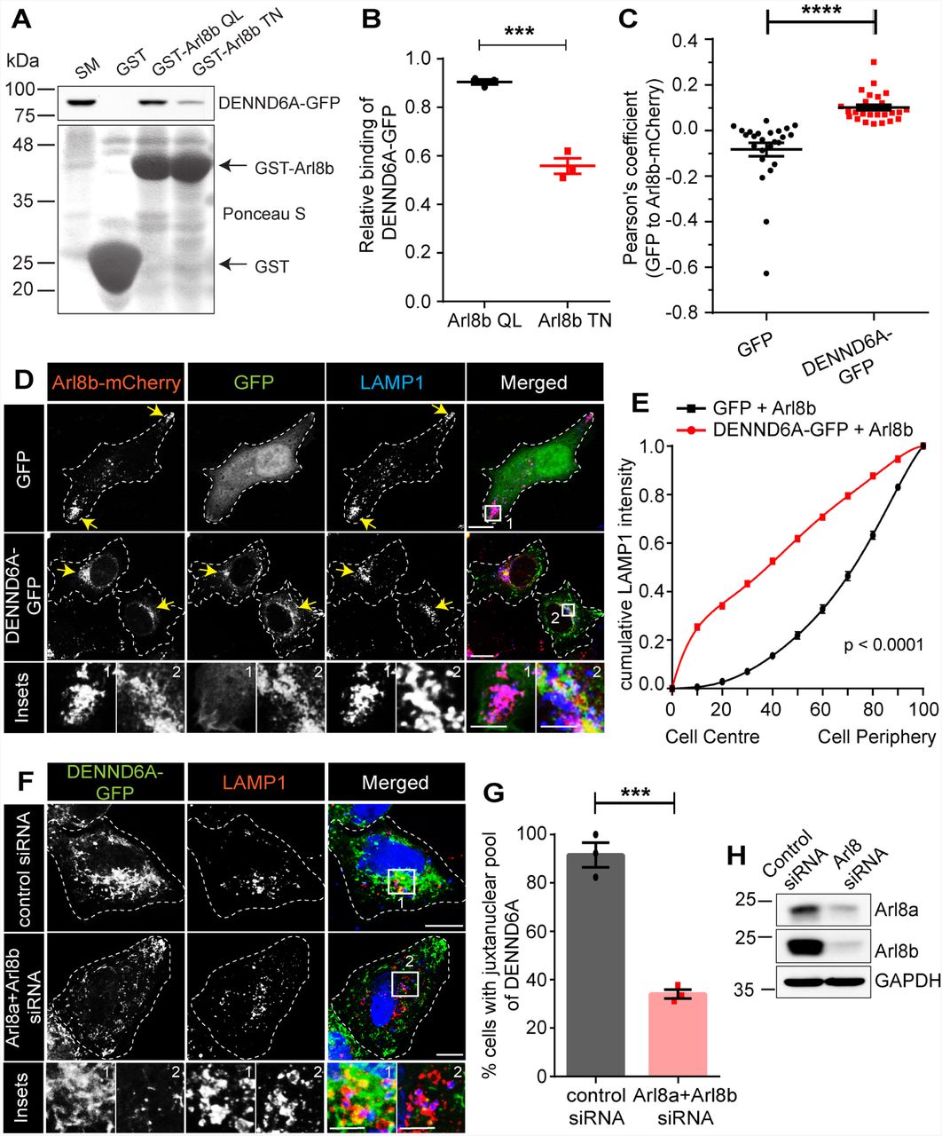
Figure Lengend Snippet: (A) Purified GST, GST-Arl8b QL and GST-Arl8b TN were incubated with lysates from HEK-293T cells expressing DENND6A-GFP. Specifically bound proteins were detected by immunoblot with anti-GFP antibody. The starting material (SM) was run in parallel to detect the total DENND6A-GFP. (B) Quantification of experiment in A ; means ± SEM; two-tailed unpaired t test (*** P ≤ 0.0005; n = 3). (C) Graphical representation of the Pearson correlation coefficient for the co-localization of GFP or DENND6A-GFP with Arl8b-mCherry from experiments performed in D ; means ± SEM; Mann-Whitney U test (**** P ≤ 0.0001; n = 25 and 26 corresponding to GFP and DENND6A-GFP). (D) HeLa cells co-expressing either GFP alone or DENND6A-GFP along with Arl8b-mCherry were fixed, stained with LAMP1 antibody and imaged using confocal microscopy (Leica SP8). The cell periphery is outlined by a white dotted line. Scale bar = 10 µm for low magnification images; 5.22 µm and 2.12 µm for high magnification images corresponding to inset 1 and 2. (E) Graphical representation of cumulative distribution of LAMP1 intensity in experiments performed in D ; mean ± SEM; extra sum of F-squares test following nonlinear regression and curve fitting; n = 25 and 26 cells, corresponding to GFP and DENND6A-GFP. (F) Control or Arl8 siRNA treated HeLa cells were transfected with DENND6A-GFP. 16 h Post transfection, cells were fixed, stained with LAMP1 antibody and imaged using confocal microscopy (Leica SP8). The cell periphery is outlined by a white dotted line. Scale bar = 10 µm for low magnification images; 3.79 µm and 4.97 µm for high magnification images corresponding to inset 1 and 2. (G) Quantification of experiment in F ; means ± SEM; two-tailed unpaired t test (*** P = 0.0004; n = 42 and 47, corresponding to control and Arl8 siRNA). (H) Immunoblot showing Arl8a and Arl8b protein levels in control and Arl8(a+b) siRNA treated HeLa cells. Immunoblot probed with anti-Arl8a, anti-Arl8b and anti-GAPDH antibodies.
Article Snippet: The following constructs were custom synthesized by SynBio technologies:
Techniques: Purification, Incubation, Expressing, Western Blot, Two Tailed Test, MANN-WHITNEY, Staining, Confocal Microscopy, Transfection

Journal: bioRxiv
Article Title: DENND6A couples Arl8b to a Rab34/RILP/dynein complex regulating retrograde lysosomal trafficking and autophagy
doi: 10.1101/2023.08.21.554162
Figure Lengend Snippet: (A) Unstarved or Earle’s Balanced Salt Solution (EBSS) starved HeLa cells were fixed, stained with LAMP1 antibody and imaged using confocal microscopy (Leica SP8). The cell periphery is outlined by a white dotted line. Scale bar = 10 µm. Yellow arrows indicate the presence of peripheral lysosomes. (B) Graphical representation of cumulative distribution of LAMP1 intensity in experiments performed in A (under starvation condition); mean ± SEM; extra sum of F-squares test following nonlinear regression and curve fitting; n = 28, 29, 30 cells, corresponding to WT, DENND6A KO1 and DENND6A KO2. (C) Immunoblot showing LC3B-II protein levels under various conditions (unstarved; EBSS starved; and EBSS starved + Bafilomycin A1 (BafA1)) Arl8a and Arl8b protein levels in control and Arl8(a+b) siRNA treated HeLa cells. Immunoblot probed with anti-LC3B-II and anti-HSC70 antibodies. (D) Quantification of experiment in F ; means ± SEM; two-way ANOVA (** P ≤ 0.0025; *** P ≤ 0.0005; **** P ≤ 0.0001; n = 3). (E) HeLa WT and DENND6A KOs cells were fixed and stained with LC3B-II antibody and DAPI. The cell periphery is outlined by a white dotted line. Scale bar = 10 µm. (F) Quantification of experiment in E ; means ± SEM; Kruskal-Wallis test (*** P = 0.0004; n = 36, 34 and 42, corresponding to WT, DENND6A KO1 and DENND6A KO2).
Article Snippet: The following constructs were custom synthesized by SynBio technologies:
Techniques: Staining, Confocal Microscopy, Western Blot

Journal: bioRxiv
Article Title: DENND6A couples Arl8b to a Rab34/RILP/dynein complex regulating retrograde lysosomal trafficking and autophagy
doi: 10.1101/2023.08.21.554162
Figure Lengend Snippet: We propose that DENND6A functions as an effector of Arl8b. The BORC complex recruits Arl8b to peripheral lysosomes, where Arl8b in turn recruits DENND6A. Once recruited, DENND6A activates Rab34, and the activated Rab34 then recruits its own effector, RILP, which is a proposed dynein adaptor. Consequently, DENND6A links Arl8b and Rab34, facilitating retrograde lysosomal transport, from the plus to minus ends of the microtubules.
Article Snippet: The following constructs were custom synthesized by SynBio technologies:
Techniques: